Breast imaging understanding how accuracy is measured when lesions are the unit of analysis review
Performance Metric
Info
- Breast imaging: understanding how accuracy is measured when lesions are the unit of analysis. Breast J. Nov-Dec 2012;18(6):557-63. doi: 10.1111/tbj.12009.
Introduction
- Breast Cancer Patients에서
- multiple lesions이 있거나 알려진 primary tumor 외의 다른 abnormal area를 찾는 경우가 있음
- e.g, Cancer 관리를 위한 mammography 이후 추가 imaging 촬영, MRI로 수술전 계획(Preoperative planning)을 세우거나 staging of disease를 정하는 것 등
- 이 경우, Radiologists가
- location of additional lesions을 정확히 찾고 이 lesions이 significant한 지 올바르게 판정하는 것이 중요함
- Significant: lesion이 malignant하거나 treatment planning을 세워야 할 정도로 malignant로 발전할 가능성이 큼(high potential)을 의미
- multiple lesions이 있거나 알려진 primary tumor 외의 다른 abnormal area를 찾는 경우가 있음
- Lesion as the unit of analysis
- 위의 경우, lesion level analysis가 필요함
- 일반적인 SN, SP, ROC curve estimates의 사용은 한계가 있음
- Purpose of the study
- 환자가 위치를 정확히 알아야 하는 multiple lesion이 있을 경우의 lesion level analysis에 대해 다룸
- lesion이 unit of analysis일 때는 일반적으로 사용하는(usual estimate) specificity나 ROC curve 사용이 어려움을 설명하고자 함
Materials and Methods
Illustrative Example
- FDG PEM(F18-2-fluoro-2-deoxy-D-glucose positron emission) MMG study
- objective: FDG PEM의 accuracy 평가
- subjects: women newly diagnosed with breast cancer(invasive or in situ)
- MRI image와 FDG PEM image를 비교
- 유방절제술(mastectomy)의 pathology information이 reference standard
Illustrative Figure
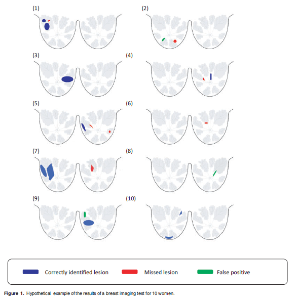
- 10 patients as an example
- it is not meant to reflect the true accuracy of any imaging test
- 9 women with significant disease, one woman without breast cancer
- 17 significant lesions (10 true positives, 7 false negatives)
- 3 false-positive findings
Methods of Analysis
- Sensitivity of a test
- the probability that the test indicates disease is present given that disease is truly present
- \(SN = P(Test + | Disease)\)
- Specificity of a test
- the probability that the test indicates there is no disease given that disease is truly absent
- \(SP = P(Test - | No Disease)\)
- Unit of Analysis가 무엇인지 중요함
Results
Patient-Level Analysis
Typical way of evaluating the accuracy of a medical test
Goal
- 질병이 있는 사람들과 없는 사람들을 test가 얼마나 잘 탐지(detect)하는 지를 평가함
2 by 2 table
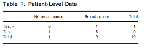
\(SN = P(Test + | Disease) = \frac{P(Test + \& Disease)}{P(Disease)} = \frac{(8/10)}{(9/10)} = 8/9 = 89%\)
\(SP = P(Test -| No\ disease) = \frac{P(Test - \& No\ disease)}{P(No\ disease)} = \frac{(0/10)}{(1/10)} = 0%\)
Breast-Level Analysis
- The breast as the unit of analysis
- 이 경우, patient level analysis에 비해 n수가 2배가 됨(each breast에 대해 평가)
- 2 by 2 Table
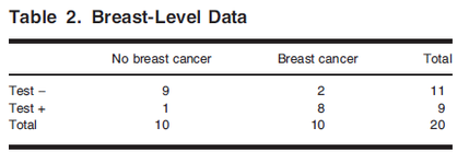
- \(SN = P(Test + | Disease) = \frac{P(Test + \& Disease)}{P(Disease)} = \frac{(8/20)}{(10/20)} = 8/10 = 80%\)
- \(SP = P(Test -| No\ disease) = \frac{P(Test - \& No\ disease)}{P(No\ disease)} = \frac{(9/20)}{(10/20)} = 90%\)
- Patient level analysis의 SP 0%가 Breast level에서는 90%가 됨
Lesion-Level Analysis
The lesion as the unit of analysis
- Figure 1에서 20 regions이 areas of interest였음
2 by 2 Table
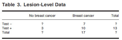
Lesion level에서는 True Negative를 정의할 수 없음
- Radiologist가 식별하지 않고 또 Pathology에서도 암이 없다고 나온 lesion을 어떻게 ‘True Negative’하다고 lesion level에서 정의할 수 있을지?
- “True Negative”라고 lesion level에서 정의할 수 있을 영역의 경우의 수가 너무 많기 때문에 위 table의 물음표(?)를 채울 명확한 방법이 없음
- (There are infinitely many possibilities for defining areas that could be marked as true negatives. The end result is that we have no obvious way to fill in this cell)
- “True Negative”라고 lesion level에서 정의할 수 있을 영역의 경우의 수가 너무 많기 때문에 위 table의 물음표(?)를 채울 명확한 방법이 없음
- 따라서 Lesion level에서는 Specificity 계산을 할 수 없음 (민감도는 가능함)
- Radiologist가 식별하지 않고 또 Pathology에서도 암이 없다고 나온 lesion을 어떻게 ‘True Negative’하다고 lesion level에서 정의할 수 있을지?
\(SN = P(Test + | Disease) = \frac{P(Test + \& Disease)}{P(Disease)} = 10/17 = 59%\)
Receiver Operating Characteristic (ROC) Curve Analysis
- Empirical ROC curve
- data are first ranked by the test result (값이 높을수록 질병이 있을 확률이 큼)
- the values above that threshold are considered positive
- For each threshold, SN and SP is calculated
- ROC Curve is the plot of SN by 1 - SP
- Lesion level에서 SP 계산을 할 수 없으므로 ROC curve도 estimate 할 수 없음
Discussion
- 예시에서 Pathology information이 available 했음
- all women in our example had a bilateral mastectomy
- breast or lesion level 평가를 위해서는 confirmed case only 등으로 study design을 조정해야함
- 한 환자가 Multiple lesions이 있는 경우
- Patient level 또는 Breast level에서는 적절히 평가할 수 없음
- the location of the lesions의 평가 또한 patient/breast level에선 고려하지 않음
- Correctly locate a lesion에 대한 test의 accuracy 평가
- LROC (Localization ROC)
- only single lesion에 대해 평가
- FROC (Free Response ROC)
- Observation은 mark-rating pairs의 형태
- Mark: location identified by a radiologist as appearing suspicious of disease
- Rating: the assessment of the probability disease is present at that location
- the fraction of identified locations that are cancerous lesions by the fraction of the identified locations that are not의 plot임
- the average number of false-positives per patient(FPP)
- Lesion level sensitivity와 함께 FPP를 보고
- Proportion of patients who have any false-positive finding도 함께 보고
- Lesion level sensitivity와 함께 FPP를 보고
- the Poisson model for the number of false positives
- the number of FPs 가 Poisson distribution을 따른다고 가정하고 Patient level에서의 Specificity를 추정함(the probability of no false-positive findings in a patient 추정에 Poisson 분포를 적용)
- The lesion-level sensitivity와 patient level specificity에 대한 joint modeling을 random effects model로 설정할 수도 있음(by Zwinderman et al.)
- LROC (Localization ROC)